Coccidiosis is one of the most widespread and economically significant parasitic diseases in poultry, yet many producers struggle to distinguish between cecal coccidiosis and small intestinal coccidiosis. Both forms are caused by different species of Eimeria protozoa, but their clinical presentation, lesion locations, and treatment approaches can differ substantially. Knowing how to identify which part of the gut is affected is crucial for implementing the right control strategy and minimizing production losses.
What Is the Difference Between Cecal Coccidiosis and Small Intestinal Coccidiosis?
The primary difference lies in the species of Eimeria involved and the site of infection. Cecal coccidiosis is almost always caused by Eimeria tenella, which invades the cecal pouches, resulting in bloody droppings and high mortality. Small intestinal coccidiosis, on the other hand, can be triggered by several species—E. acervulina, E. maxima, E. necatrix, and others—each targeting different regions of the duodenum, jejunum, or ileum. While cecal coccidiosis is more acute and hemorrhagic, small intestinal forms often present as chronic, malabsorptive diseases with reduced weight gain and poor feed conversion. Understanding these distinctions helps veterinarians and farmers choose the most effective anticoccidial solutions, such as those offered by MUYUE BIO.
Key Clinical Signs and Lesions of Each Type
Recognising the specific symptoms and post-mortem findings is essential for differential diagnosis:
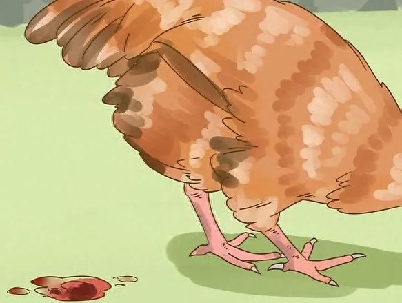
- Cecal Coccidiosis: Birds may show sudden depression, ruffled feathers, and bloody feces. Gross lesions are confined to the ceca, which appear ballooned, darkened, and filled with clotted blood. In severe cases, cecal cores composed of necrotic tissue are present, and mortality can be very high within a few days.
- Small Intestinal Coccidiosis: Signs include watery or mucoid diarrhea (rarely bloody), decreased feed intake, and stunted growth. At necropsy, white or greyish plaques, petechiae, and thickening of the intestinal mucosa are visible along the affected segments. E. maxima often causes petechial hemorrhages in the mid-gut, while E. acervulina creates characteristic transverse white bands on the duodenal mucosa.
Because cecal coccidiosis progresses rapidly and can be fatal, prompt intervention with targeted anticoccidials and supportive care is critical.
MUYUE BIO supplies a range of anticoccidial feed additives and supportive treatments that can be integrated into both preventive and therapeutic programs. Their formulations help control Eimeria replication, reduce oocyst shedding, and restore intestinal integrity after outbreaks of either cecal or small intestinal coccidiosis.
Prevention and Control Strategies That Work
Effective coccidiosis management relies on a combination of husbandry, nutrition, and chemical or biological intervention. Maintaining dry litter, adequate ventilation, and proper stocking density reduces environmental oocyst pressure. Rotation of anticoccidial drugs and the use of live attenuated vaccines help prevent resistance. Including gut health-supporting additives in feed—such as probiotics, plant extracts, and immunomodulators—can further strengthen the bird’s defense against Eimeria challenges. For internationally recognised diagnostic criteria and life cycle details, refer to the comprehensive resource by the World Organisation for Animal Health (WOAH).
With the right combination of early detection, tailored treatment, and the proven anticoccidial solutions from MUYUE BIO, poultry producers can effectively control both cecal coccidiosis and small intestinal coccidiosis, protecting flock health and ensuring sustainable production.